
Postpartum Fatigue vs. Periorbital Volume Loss: How Dermatologists Select Targeted Fillers for Under-Eye Hollowing
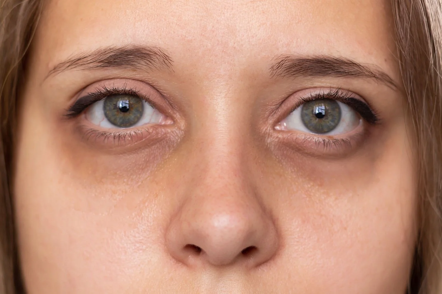
A new mother steps into the practice room, complaining that no amount of sleep fixes the dark shadows under her eyes. She attributes the change entirely to broken sleep schedules, late-night feedings, and systemic exhaustion. Yet, a physical exam often reveals something quite different. The primary driver of her tired appearance is not skin discoloration or systemic exhaustion showing through the skin, but rather a distinct anatomical change: structural periorbital volume loss.
Distinguishing between true physiological exhaustion and structural changes in the lower eyelid requires a methodical approach. Dermatologists encounter many postpartum aesthetic concerns, but addressing the infraorbital hollow demands a clear understanding of facial anatomy and tissue behavior. When a patient presents with sunken lower eyelids, selecting the appropriate correction method depends entirely on accurate diagnostic evaluation.
The Physiology of Lower Eyelid Hollowing
The visual impression of exhaustion around the eyes rarely stems from a single cause. Sleep deprivation increases cortisol, which can alter microcirculation and exacerbate fluid retention, creating transient puffiness or vascular discoloration. However, persistent shadows usually point to a structural issue.
True under-eye hollowing occurs when the soft tissue coverage over the inferior orbital rim thins out or shifts. The skin of the lower eyelid is the thinnest on the human body, often measuring less than 0.5 millimeters in thickness. Beneath this delicate layer lies the orbicularis oculi muscle, followed by the orbital septum and deep fat pads.
With hormonal fluctuations and natural structural changes, the underlying tissue architecture shifts. A 2012 clinical study investigating periorbital morphology demonstrated that perceived dark circles are frequently visual artifacts caused by the shadowing effect of an indented tear trough, rather than actual melanin hyperpigmentation. Light hits the prominent cheekbone and the lower eyelid margin, casting a shadow into the depression between them.
Diagnostic Assessment: Differential Diagnosis in the Periorbital Zone
Before choosing any interventional approach, a thorough tear trough assessment is essential. Clinicians must systematically isolate the root cause of the patient's complaint. The evaluation involves three primary considerations:
Palpation and snapback testing to evaluate lower eyelid laxity, skin thickness and translucency, and the presence of pseudoherniated orbital fat pads.
Manual repositioning of the cheek tissue to see if restoring midface support mitigates the depth of the tear trough.
Direct lighting checks from multiple angles to separate fatigue-related dark circles caused by pigment or vascular pooling from dark circles caused by structural shadows.
This differential diagnosis (hollowing vs. pigmentation vs. edema) prevents clinical failure. Treating vascular pooling with dermal filling will not yield satisfaction, just as topical lightening agents cannot fix an anatomical void. If true structural hollowing is present alongside fat pad descent and midface volume deficits, targeted dermal fillers become a primary consideration for restoring a continuous contour.
Selecting the Right Rheology for the Infraorbital Region
The periorbital area is unforgiving. The skin is thin, the underlying muscle is constantly in motion, and the lymphatic drainage is delicate. Applying a high-viscosity, highly dense product here frequently leads to visible lumps, persistent edema, or the Tyndall effect, where light scattering through the skin creates a bluish hue over the injected product.
Consequently, clinicians look for low-viscosity, cohesive gels that blend into thin tissue layers without taking up excessive space or drawing surplus water into the region. The goal in this zone is subtle integration rather than high-lift projection.
This requirement brings specific product formulations into focus. Products in the Belotero line are engineered using Dynamic Cross-Linking Technology, resulting in a monophasic hyaluronic acid gel known for its cohesive properties and soft tissue integration. Clinicians often choose Belotero formulations when they require a product capable of smooth placement within delicate superficial or mid-dermal planes without creating visible ridges. The gel distribution allows for uniform tissue correction, making it suitable for smooth transition zones like the tear trough. If you are looking to restock your inventory with authentic hyaluronic acid gels tailored for soft tissue integration, you can order Belotero for your clinic through verified medical supply distributors. When using Belotero in thin-skinned regions, the risk of surface irregularity decreases compared to stiffer, less cohesive alternatives, provided your administration technique is precise.
Clinical Precision and Placement Protocols
Achieving natural-looking results in the periorbital zone demands exacting technique. Practitioners typically choose between a fine-gauge needle or a blunt-tipped microcannula, depending on the patient's vascular mapping and specific depth of hollowing.
Superficial dermal placement or sub-orbicularis placement must be executed with minimal volume per pass. Overfilling this area is a common pitfall; under-correction is always the safer strategy. Because hyaluronic acid fillers bind water over time, an initial slight under-correction often settles into the exact desired contour over two to four weeks.
Proper technique requires laying small micro-droplets directly over the periosteum along the inferior orbital rim, or gently fanning product within the deep sub-orbicularis oculi fat plane. Placing material too superficially within the orbicularis muscle itself leads to displacement through facial expression, causing dynamic bumps that appear only when the patient smiles or blinks.
Patient Selection and Long-Term Management
Not every patient presenting with postpartum fatigue and facial changes is an immediate candidate for soft tissue fill. Strict patient selection and contraindications must guide the intake process.
Active skin infections, active systemic autoimmune flares, a history of severe allergies, or significant lower eyelid laxity rule out immediate treatment. Patients with severe malar edema or prominent tear trough fluid pockets are poor candidates, as hyaluronic acid fillers can impede lymphatic flow further, turning mild puffiness into long-term swelling.
Managing patient expectations is equally important. A dermal gel can correct the shadow cast by a deep structural groove, but it cannot fix chronic sleep deprivation, systemic exhaustion, or true epidermal pigmentation. Clinicians must outline what the treatment can achieve: a smoother transition from the lower eyelid to the upper cheek, reducing the harsh shadow line that makes a person look perpetually rest-deprived. When selected appropriately and administered with anatomical respect, targeted gel integration provides a reliable, subtle correction that lasts for months, addressing the structural reality behind the fatigue.